How to make a temporary slide
Cells are tiny, so we use microscopes to see their details.
A slide is a thin piece of glass used to hold objects which are examined under a microscope.
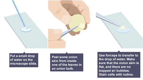
Plant cells:
Peel a thin layer of cells from an onion.
Place on a slide.
Add a drop of iodine (a chemical stain).
Gently lower a coverslip to avoid air bubbles and prevent drying.
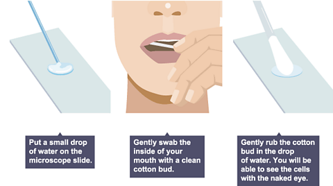
Animal cells:
Collect cheek cells with a cotton bud.
Smear onto a slide.
Add methylene blue (a chemical stain).
Gently lower a coverslip to avoid air bubbles.
Chemical stains highlight cell parts making them easier to see. It’s temporary as the cells are not preserved so will rot.
Observing and recording
Using a light microscope
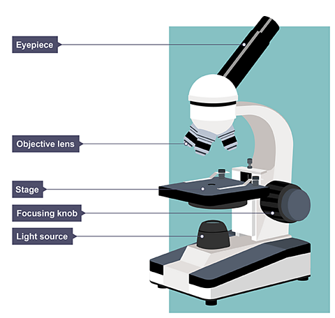
When using a light microscope, start with the low power objective lens as the field of view will be wider.
This will increase the number of cells you are able to see, making it easier to locate cells.
Once centred, switch to a higher power lens for detailed viewing.
The lens is very close to the slide so be careful to avoid damaging it.
Total magnification = eyepiece magnification × objective lens magnification.
| Objective lens | Eyepiece magnification | Total magnification | |
|---|---|---|---|
| Low Power | 4X | 10X | 40X |
| Medium Power | 10X | 10X | 100X |
| High Power | 40X | 10X | 400X |
Drawing cell structures:
A good biological drawing should be:
Drawn in pencil with firm, continuous lines (no sketching).
Large and proportionate to the observed cell.
Clearly labelled.
Given a title with magnification or size.
Size and magnification
Magnification can be worked out from a photograph or drawing.
You need to measure using The standard units of measurement used in biology to describe quantities like length, mass, and time. - millimetre (mm) or micrometre (µm). Ensure the same unit is used throughout the calculation.
1 meter (m) = 1,000 millimetres (mm)
1 millimetre (mm) = 1,000 micrometres (µm)
You can convert between these units by multiplying or dividing by 1,000.
You need to determine the size of biological specimens by the following methods:
1. Estimation:
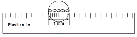
a) Place a clear plastic ruler under the microscope.
b) Measure the total width of several cells (e.g., 5 cells).
c) Divide the total length by the number of cells: 1 mm / 5 cells = 0.2 mm per cell
d) Each cell is approximately 0.2 mm wide.
magnification = size of image ÷ size of real object
The above equation can be rearranged to calculate the actual length of the cell and the magnification used as well as the size of the image.
Size of Image = Actual Size x Magnification
Actual Size = Size of Image ÷ Magnification
The triangle can be used to help remember this formula.
2. Calculation using Magnification:
a) Use a ruler to measure the size of the specimen in the image (eg 10 mm).
b) Find the Magnification: (eg 100x). This information is often provided by the microscope or drawing.
c) Apply the formula:
\(\text{Actual size} = \frac{\text{Size of image}}{\text{Magnification}}\)
For example, if the image size is 10mm and the magnification is 100x:
\(\text{Actual size} = \frac{\text{100mm}}{\text{100}}\) = 0.1mm = 100µm
This means the actual size of the specimen is 100 micrometers.
Higher Tier only:
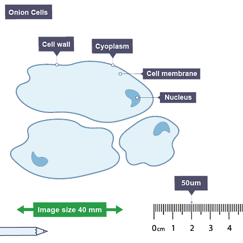
Using a Scale Bar to calculate magnification:
a) Locate the scale bar on the image - the line drawn which has a label showing the actual length of the bar before being magnified eg 50µm.
b) Measure the length of the scale bar using a ruler in mm eg 2cm = 20mm.
c) Convert to micrometres (multiply by 1000) eg 20mm x 1000 = 2000 µm.
d) Calculate the magnification - scale bar length divided by actual scale bar length (written on the scale bar) eg 2000 µm / 50µm = X 40
Electron microscopes
Increasing magnification beyond a certain point does not necessarily improve image detail.
Electron microscopes have a much higher resolution than light microscopes, allowing us to see more detail of cell structures.
They use electron beams which are much smaller than light waves, to achieve a high resolution.
This increased resolution has improved our understanding of cell structure by:
Providing a clearer and more detailed view of cells (eg ribosomes)
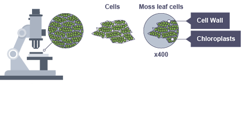
Revealing structures of smaller organelles (eg mitochondria, chloroplast )
Quiz time!
Watch: The power of the microscope
More on Cells
Find out more by working through a topic
- count2 of 3

- count3 of 3
